Part 2: Anatomical Alignment
To make judgements about biomechanical features of a foot on X-ray, dorsoplantar and lateral weight bearing radiographs are obtained in normal angle and base of gait to provide an accurate representation of the foot in its functional position.
Dorsoplantar view
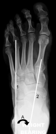
Line 1 = longitudinal axis of hindfoot
Line 2 = midline axis of talus
A = talocalcaneal angle
Longitudinal axis of the hindfoot
A line parallel to the distal lateral border of the calcaneus.
Normal: parallel to the long axis of the fourth metatarsal.
Talo-calcaneal angle
Measured between the longitudinal axis of the hindfoot and a line along the midline axis of the talus.
Normal: 15-35°
Hindfoot varus
Smaller talo-calcaneal angle means the calcaneus is directed closer to the midline and there is hindfoot varus or supination with increased superimposition of the talus on the calcaneus.
Hindfoot valgus
Larger talo-calcaneal angle means the calcaneus is directed further from the midline and there is hindfoot valgus or pronation.
Forefoot varus and valgus
Often assessed as a relationship between the talus and the first metatarsal.
Normal: midline axis of the talus goes through the base of the first metatarsal.
If the axis of the talus lies lateral to the first metatarsal, the metatarsal is directed closer to the midline and there is forefoot varus or supination (also referred to as inversion). The bases of the metatarsals tend to converge more than normal and the proximal metatarsals are superimposed.
If the axis of the talus lies medial to the first metatarsal, the metatarsal is directed further from the midline and there is forefoot valgus or pronation (also referred to as eversion). The metatarsals are less convergent than normal, more parallel with less overlap.
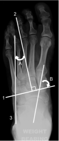
Line 1 = bisection of lesser tarsus
Line 2 = longitudinal axis of lesser tarsus
Line 3 = longitudinal axis of hindfoot
A = lesser tarsus abduction angle
B = talonavicular angle
Longitudinal axis of the lesser tarsus
Perpendicular to a line that transects the lesser tarsus which extends across the tarsus from halfway between the medial aspect of the talo-navicular joint and the medial aspect of the first tarso-metatarsal joint to halfway between the lateral aspect of the calcaneo-cuboid joint and the lateral aspect of the fifth tarso-metatarsal joint.
Lesser tarsus abduction angle
Between the longitudinal axis of the lesser tarsus and the longitudinal axis of the hindfoot.
This angle increases with pronation and decreases with supination.
Talo-navicular angle
Between the midline axis of the talus and the bisection of the lesser tarsus.
Normal: 60-80°
This angle is >80° in the supinated foot and <60° in the
pronated foot.
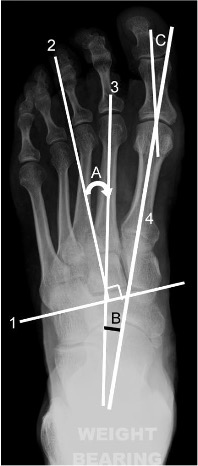
Line 1 = bisection of lesser tarsus
Line 2 = longitudinal axis of lesser tarsus
Line 3 = longitudinal axis of metatarsus
Line 4 = midline axis of first metatarsal
A = metatarsus adduction angle
B = metatarsus primus adductus (or intermetatarsal) angle
C = hallux valgus angle
Longitudinal axis of the metatarsus
A longitudinal bisection of the second metatarsal.
Metatarsus adduction angle
Between the longitudinal axis of the metatarsus and the longitudinal axis of the lesser tarsus.
Normal: <15°
A greater angle indicates metatarsus adductus or deviation of the forefoot to the midline. Severe metatarsus adductus may be associated with hindfoot valgus (e.g. skew foot).
Hallux valgus angle
Between the longitudinal bisection of the first proximal phalanx and first metatarsal.
Normal: <15°
Mild: 16-25°
Moderate: 26-35°
Severe: >35°
Lateral view
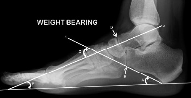
Line 1 = calcaneal inclination axis
Line 2 = midline axis of talus
A = calcaneal inclination angle
B = talar declination angle
C = lateral talocalcaneal angle
D & E indicate the cyma line
Calcaneal inclination axis
A line along the inferior surface of the calcaneus connecting the most inferior point of the calcaneal tuberosity with the most distal inferior point of the calcaneus at the calcaneo-cuboid joint.
Calcaneal inclination angle
Between the calcaneal inclination axis and the supporting surface, reflecting the height of the foot framework.
Measurements of 10-20° are considered low, 20-30° medium and >30° high. Angle varies with foot pronation or supination and clinical judgement is important.
Talar declination angle
Between a bisection through the body and neck of the talus and the supporting surface.
Normal: ~21°
Pes cavus and pes planus
Meary’s angle is formed from the midline talar axis and the first metatarsal shaft axis.
Normal: 180°; i.e. should be a straight line (on DP and lateral views).
If the first metatarsal axis extends downwards compared to the talar axis (ie the first metatarsal is steeper in relation to the horizontal), the talar declination angle decreases and the line falls above the first metatarsal. This indicates a high longitudinal foot arch and a pes cavus foot.
If the talar axis extends downwards compared to the first metatarsal axis, the talar declination angle increases and the line falls below the first metatarsal. This indicates a low to flat longitudinal foot arch and a pes planus foot.
Another method for assessing cavus or planus is measuring the angle of the longitudinal arch between the calcaneal inclination axis and a line which extends along the undersurface of the fifth metatarsal (normal 150-170°).
Cavus is present with an angle of less than 150°.
In planus the angle is increased approaching 180°.
Lateral talo-calcaneal angle
Between the calcaneal inclination axis and the midline axis of the talus.
Normal: 35-50°
Bohler’s and Gissane's angles
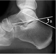
B = Bohler’s angle
Between a line from highest point of calcaneal anterior process to highest point of posterior facet and a line tangential to the posterior superior margin of the calcaneus.
Normal: 20-40°
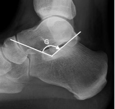
G = Gissane angle
Between the downward (body and anterior calcaneal process) and upward (posterior articular facet) slopes of the superior calcaneal surface.
Normal: 120-145°
Calcaneus and equinus
Normally the undersurface of the calcaneus is higher anteriorly than posteriorly.
If the anterior calcaneus extends lower than the posterior, or if they are at the same level, an equinus calcaneus is present.
If the anterior calcaneus extends more upward than normal, above the rest of the upper surface of the bone, a calcaneus calcaneus is present.
The talo-navicular and calcaneo-cuboid joints together produce a superimposition on a lateral X-ray known as the cyma line. These curved joints together form a ‘lazy S’ as an intact curve in a normal foot. In a pronated foot the ‘S’ becomes broken as the talo-navicular joint moves anterior and plantar to the calcaneo-cuboid joint and the reverse occurs in a supinated foot