Acute Diabetic Foot
Introduction
Patients with diabetes are prone to develop foot complications due to associated peripheral neuropathy, peripheral vascular disease, and the systemic manifestations of diabetes. The peripheral nervous system normally contributes to the early warning response and neuropathy diminishes the body’s ability to respond appropriately to stressful insults.
The spectrum of clinical manifestations of foot complications in diabetes is termed as diabetic foot disease. Acute manifestations of diabetic foot disease that are generally managed by an orthopaedic surgeon include an acute Charcot foot and acute severe infection (Diabetic Foot Attack)
Acute Charcot Foot
Charcot neuroarthropathy (CN) is one of the most devastating complications of diabetic peripheral neuropathy. Acute (active) CN is often triggered by minor trauma to the foot and results in the development of an uncontrolled inflammatory response. This leads to bone fragmentation, fractures, and joint dislocations. Due to the presence of neuropathy, pain sensation is reduced, and patients continue to weight bear, resulting in further bone fragmentation and deformation.
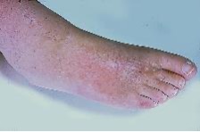
The patient typically presents with a painless, red, warm, swollen foot with (perhaps) a history of minor trauma. Examination reveals a temperature ≥ 2°C higher than the normal foot. The pedal pulses are usually palpable. There may be deformity with crepitus on palpation due to the underlying bone fragmentation, particularly with delayed presentation.
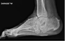
Differential diagnoses include cellulitis, deep infection, DVT, and gout; appropriate investigations may be considered to rule out these pathologies. Plain radiographs may initially appear normal but show bone fractures and joint dislocations in late presentations. MRI is extremely useful in the diagnosis of early acute Charcot (but can be difficult to differentiate from infection).
Based on plain radiographs, CN is classified using the Eichenholtz classification as follows:
- Stage 1: destruction / fragmentation
- Stage 2: coalescence
- Stage 3: consolidation
Stage 0 was recently added in which x-rays are normal, but MRI shows evidence of bone oedema and subchondral fractures.
Coalescence occurs as the acute and uncontrolled inflammation and oedema settles, along with absorption of fine bone debris. This now becomes inactive (or chronic) CN. Consolidation occurs once oedema fades and the bone remodels.
Chronic CN can present with various degrees of foot and ankle deformities, ulceration over bony prominences, and osteomyelitis in some cases.
Based on the location of CN changes, Sanders and Fryberg have classified 5 anatomical patterns:
- MTP joints
- TMT joints
- Tarsal joints
- Ankle joint
- Body of calcaneum
Management of Acute Charcot
Non-operative management
This involves the immediate offloading of the leg in a total contact cast (TCC) and protected weight-bearing. TCC allows:
- even distribution of plantar pressures across the sole of the foot and load transfer to the leg
- reduction of oedema in the lower leg and ankle
- optimisation of mechanical and biological environment
- helps to resolve the inflammatory phase, minimising bone fragmentation
- helps to prevent progression to a major deformity
- duration between 3-12 months
TCC is continued until the inflammatory response settles – as noted by resolution of swelling and normalisation of temperature – followed by gradual return to normal weight-bearing in a brace, custom-made shoes or a CROW (Charcot Restraint Orthotic Walker).
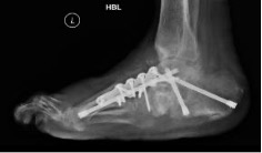
Despite adequate offloading, deformities may progress with bony instability resulting in ulceration and infection. Thus, the foot may become at risk of a major amputation. To achieve functional limb salvage, such presentations warrant immediate surgical stabilisation, even during Eichenholtz stage 1. This management is best delivered by a multi-disciplinary diabetic foot team. The patient is admitted immediately for continuous leg elevation and TCC offloading to reduce swelling and normalise foot temperature. Surgical stabilisation of the Charcot foot is performed using the principles of durable, long-segment, rigid internal fixation with optimal bone opposition. Recent small series studies have shown improved results of surgical reconstruction of active CN. Fine-wire circular frames can also be used for early stabilisation and deformity correction.
Diabetic Foot Attack
The commonest manifestation of foot infection in the presence of peripheral neuropathy is an infected diabetic foot ulcer. Classical symptoms and signs of infection are often delayed as their expression is dependent on an intact peripheral nervous system. However, most diabetic foot ulcers respond favourably to bedside podiatric debridement, offloading in a TCC and administration of a course of deep tissue sample, culture-specific, oral antibiotics. Occasionally, some infections progress rapidly along the tissue planes with signs of spreading cellulitis, tissue necrosis and systemic inflammatory response. Such infections can be limb-threatening without timely intervention and have been labelled as ‘diabetic foot attack’. The management of diabetic foot attack is ideally delivered by a multi-disciplinary diabetic foot team using a structured approach. The foot and ankle surgeon is expected to take the lead in the surgical component of management.
Principles of management of diabetic foot attack
DFA is primarily managed surgically by resection of all infected and necrotic tissue; achieve infection eradication and predictable ulcer healing. The principles of management are:
- Rapid diagnosis; the concept of ‘time is tissue’ is applicable
- Clinical presentation, blood investigations (raised CRP), urgent MRI +/- vascular studies
- Identifying pathogens: culture of deep tissue samples, US-guided aspiration of deep collection
- Aggressive and targeted IV antibiotic therapy
- Emergent and radical surgical debridement of all infected and necrotic tissue
- surgical exploration along tendon sheaths and tissue planes
- local antibiotic-eluding calcium sulphate preparation may be used to pack tissue voids created by surgical debridement
- repeat debridement if required
Non-healing ulcers or recurrence of infection are often due to inadequate surgical resection of the infected and non-viable tissues. Any soft tissue defect following debridement is best managed with off-loading in a bi-valved TCC, negative pressure wound therapy and, later, split skin graft coverage.
MCQs
- Which of the following clinical feature is NOT indicative of active Charcot foot?
- Swelling
- Redness
- Local warmth
- Absent foot pulses
- Absence of pain
- Which of the following statement is applicable for Diabetic Foot Attack?
- This can be effectively managed non-operatively
- Extensive investigations and prolonged monitoring are required to achieve a diagnosis
- Aggressive surgical debridement of all infected and necrotic tissues is essential to prevent recurrence of infection
- This should be treated by the vascular team only
- Superficial wound swabs always identify the true pathogens
References
Frykberg R, Wukich D, Kavarthapu V, Zgonis T, Dalla Paola L. Surgery for the diabetic foot: a key component of care. Diabetes Metab Res Rev. 2019;e3251.
Simon SR, Tejwani SG, Wilson DL, Santner TJ, Denniston NL. Arthrodesis as an early alternative to nonoperative management of Charcot arthropathy of the diabetic foot. J Bone Joint Surg Am. 2000;82-A(7):939-50.
Vas PRJ, Edmonds M, Kavarthapu V, Rashid, H, Ahluwalia, R, Pankhurst, C, Papanas, N. The Diabetic Foot Attack: "'Tis Too Late to Retreat!". Int J Low Extrem Wounds. 2018;17(1):7-13.doi:10.1177/1534734618755582