Posterior Malleolus Fractures
Introduction
- Posterior malleolus fractures (PMF) are observed in approximately 15-44% of ankle fractures
- Historically, the size of the PMF on lateral ankle radiographs was used to determined whether it should be managed operatively
- Systematic reviews have shown that the size of the PMF fragment is not a predictor of outcome
- Functional outcomes are generally poor historically
Investigation
Plain radiographs are not reliable enough to provide sufficient information about PMF fragments
CT scan is the preferred test:
- Fracture configuration
- ‘Die-punch’ fragments
- Intra-articular fragments
Classification
Figure 1: Mason & Molloy (2017)
.jpg?ver=Tl2JRqTzeEdMORp0nj6_Yg%3d%3d)
.jpg?ver=Tl2JRqTzeEdMORp0nj6_Yg%3d%3d)
- Other classification system include:
- Haraguchi
- Bartoniček/Rammelt
Fixation Technique
Direct Approach
- Level 1 evidence shows significant improvement in anatomical reduction and functional outcome with the direct approach
- Primary wound healing was achieved in all cases, with no infection, when medial posterior medial and posterior lateral approach done together
|
Classification (Mason & Molloy)
|
Treatment
|
Approach
|
|
1
|
Syndesmotic Fixation
|
Direct lateral (fibular)
|
|
2A
|
ORIF
|
Postero-lateral (PL)
|
|
2B
|
ORIF Postero-medial Fragment First
|
Postero-lateral + Medial Posterior Medial (MPM)
or Postero-medial (PM)
|
|
3
|
ORIF
|
Posterior Medial
|
Table 1: Liverpool algorithm for direct fixation of posterior malleolar fracture
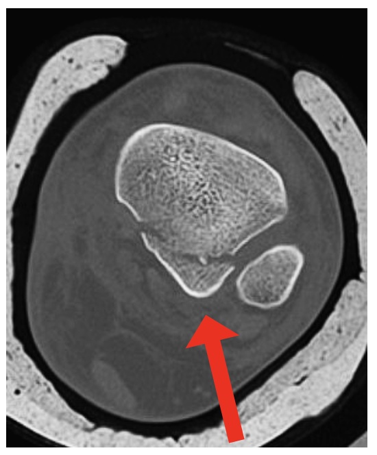
Figure 2 Posterolateral (PL) approach:
- Incision mid-way between Achilles tendon and fibula
- Watch out for sural nerve
- Approach between FHL and peroneals (true inter-nervous plane)
- Avoid damaging the PITFL - needs preserving to maintain posterior syndemosis stability
- Can combine with separate, standard medial malleolus approach
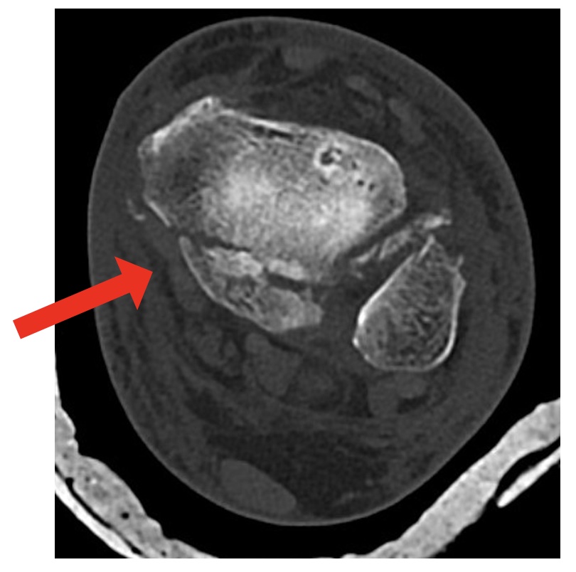
Figure 3: Medial postero-medial (MPM) approach
- Incision behind, and then curving inferior to, medial malleolus
- Approach onto tendon of FDL - can be mobilsed (+/- tib post) to visualise fracture(s)
- Look for NV bundle - keep it lateral
- With 2B patterns, reduce and fix postero-medial fragment before postero-lateral fragment
- Can fix medial malleolus through same incision
- Can combine with separate, standard lateral approach for fibula / syndesmosis fixation
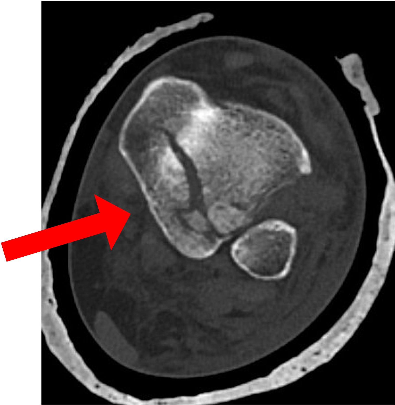
Figure 4: Postermedial (PM) approach
- Incision closer to Achilles tendon
- Look for NV bundle - keep it medial
- Good view of majority of posterior face of tibia for stable fixation
- Can combine with separate, standard lateral and/or medial malleolus incisions
Indirect Approach
- Screw directed from an anterior to posterior direction (“AP” Screw)
- Concerns:
- Screw displaces the PMF
- Soft tissue / bony entrapment in the fracture site
- High incidence of injury to anterior structures (esp with percutaneous approach)
Outcomes
- Good functional outcome equivalent to bimalleolar fractures with fragment specific fixation
- Fracture morphology dictates functional outcomes (not fragment size)
- Rotational pilons did worse
- Reduction is key for posterior malleolar fractures fixation
- Post-operative articular step of the posterior malleolus leads to higher incidence of post-traumatic osteoarthritis
Syndesmotic Injury
- Mason and Molloy:
- Type 1 fractures ~ 100% have syndesmotic injury
- Type 2 fractures ~ 49% have syndesmotic injury
- Type 3 fractures ~ 20% have syndesmotic injury
- The insertion of the posterior inferior tibio-fibular ligament (PITFL) is broad at the posterior aspect of the tibia; therefore, not all M&M Type 2 and 3 fractures are associated with syndesmotic injury
- PMF fracture fixation reduces the need for syndesmotic stabilisation
Die-punch Fragments Associated with Posterior Malleolar Fractures
- Die-punch fragment size may not impact clinical and functional outcome but may contribute to post-traumatic arthritis
- Intra-articular impacted fragment can potentially lead to articular malreduction and post-traumatic arthritis
- Removing die-punch fragments can be challenging, due to poor visualisation via the posterior approaches described; access can be achieved through the Weber B fibular fracture, or through the apex of the PMF fragment
Controversy in Posterior Malleolar Fracture Fixation
- Trimalleolar fractures have a worse outcome than bimalleolar fractures due to the chondral damage sustained
- Direct fixation of PMF fractures have a reported complication rate of around 17-20% including infection, sural nerve neuropraxia, loss of reduction, mal-reduction (28%) early arthritis and major limb amputation
Summary
- Obtain a CT scan preoperatively when possible
- Fragment specific fixation has shown improve functional outcomes
- Fracture morphology and articular step are better predictors of outcome than the size of posterior malleolar fracture
- Next steps include better techniques to manage the articular surface injury
Video Resources
https://www.youtube.com/watch?v=Y55YV2rHfkw – Lyndon Mason Hunterian Lecture 2020
https://www.youtube.com/watch?v=u23nRei9dsc – Andy Molloy BOFAS Lectures of Distinction 2020
https://www.youtube.com/watch?v=0wpsGl4fdBQ – Lyndon Mason Edinburgh International Trauma Symposium
https://www.youtube.com/watch?v=DrTDEMzWOWM – Lyndon Mason – Orthohubxyz Posterior Malleolar Approaches 2020
References
Verghage et al. When and how to operate the posterior malleolus fragment in trimalleolar fractures: a systematic literature review. Arch Orthop Trauma Surg. 2018 Sept;138(9):1213-1222
Gandham et al. Posterior malleolar fractures: A CT guided incision analysis. Foot (Edinb). 2020 Jun;43:101662
Mason et al. Pathoanatomy and Associated Injuries of Posterior Malleolus Fracture of the Ankle. Foot Ankle Int. 2017 Nov;38(11):1229-1235
Vidovic et al. Posterior Fragment in Ankle Fractures: anteroposterior vs posteroanterior fixation. Injury. 2017 Nov;48 Suppl 5:SS65-S69
Yang et al. Combined Posteromedial and Posterolateral approaches for 2-Part Posterior Malleolar Fracture Fixation. Foot Ankle Int. 2020 Oct:41(10):1234-1239
Gandham et al. Posterior malleolar fractures: A CT guided incision analysis. Foot (Edinb). 2020 Jun;43:101662
Philpott et al. Posterior Approaches to the ankle – an analysis of 3 approaches for access to the posterior malleolar fracture. Foot (Edinb). 2020 Dec;45:101725
Mason et al. Posterior Malleolar Ankle Fractures: An Effort at Improving Outcomes. JBJS Open Access. 2019 Jun 7;4(2):e0058
Blom et al. Posterior malleolar ankle fractures. Bone Joint J. 2020 Sept;102-B(9):1229-1241